
Study Reference
Title
Comparative analysis of the use of autologous exosomes and platelet-derived growth factors in women with premature ovarian insufficiency and infertility: A prospective, randomized, observational, analytical study
Journal
Regenerative Therapy (2025)
Statement
This summary is based on the original publication and includes application-oriented discussion for educational and academic reference purposes only. It is not intended as medical advice.
Summary
1. Introduction: The Biological Mystery of Age 35
In the field of reproductive longevity, age 35 is frequently cited as the “biological cliff.” For decades, the prevailing clinical narrative has focused almost exclusively on the depletion of the primordial follicle pool—the idea that women are simply “running out of eggs.” However, as a longevity strategist, I look deeper into the “soil” rather than just the “seeds.”
Emerging evidence suggests that ovarian aging is driven by a profound physical transformation of the ovarian environment itself. We are beginning to understand that the ovary doesn’t just retire; it becomes structurally compromised. This post explores a groundbreaking shift in “Ovarian Rejuvenation” using autologous exosomes—a method that seeks to repair the biological infrastructure and potentially rewind the clock on fertility.
2. The Hidden Cost of Ovulation: 450 Monthly Scars
Every month, the process of ovulation—the hallmark of a healthy reproductive system—exacts a cumulative physical price. When a follicle ruptures to release an oocyte, it causes microscopic trauma to the ovarian cortex. This repeated wounding leads to a condition known as stroma fibrosis (or “stroma fabulosis”).
The math of this decline is stark. With an average of 13 ovulations per year over roughly 35 years of menstruation, a woman reaching the age of 35 has sustained approximately 450 physical scars on her ovaries. By age 37, this accumulation of fibrotic, collagen-heavy tissue fundamentally alters the organ’s landscape.
This is the great biological irony: the very process required for fertility eventually creates a physical barrier to it. These “450 scars” impede blood circulation and nutrient delivery, creating a state of metabolic stagnation. In a young ovary, the blood supply is robust; in a scarred ovary, the dense fibrosis acts as a wall, leading to localized hypoxia and preventing the remaining follicles from receiving the signals they need to mature.
3. The Ovarian Engine: Why Mitochondria Matter
To appreciate the impact of this physical decay, we must understand the energy requirements of the oocyte. The ovary is a high-performance organ, ranking second only to the brain in mitochondrial density. Mitochondria are the “engines” producing adenosine triphosphate (ATP), the fuel required for the intricate dance of cell division.
As the source context highlights:
“In the process of egg formation, it must first undergo mitosis and then meiosis. During this, a lot of energy is required.”
When stroma fibrosis limits circulation, it starves the mitochondria of oxygen. This hypoxia leads to an energy deficit that directly correlates with chromosomal abnormalities. Without sufficient ATP, the cellular machinery lacks the power to divide chromosomes accurately. This results in deletions, duplications, and trisomies (such as Trisomy 21/Down Syndrome). While many of these errors lead to early pregnancy loss, others may not be detected until week 16 of gestation, leading to difficult clinical decisions. Repairing the “engine” by addressing the environment is therefore critical for genetic integrity.
4. The 2025 Breakthrough Study: Exosomes vs. PRP vs. Placebo
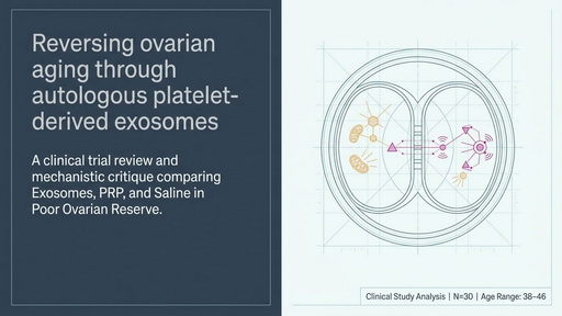
A pivotal clinical study published in November 2025 offers a glimpse into how we might dismantle this fibrotic barrier. The study followed 30 participants, aged 38 to 46 (average age 41.2), all characterized by low ovarian reserve (AMH < 1.0) and elevated FSH levels.
The trial compared three intervention groups, with participants receiving 2cc injections into each ovary once a month for four consecutive months:
- Autologous Exosomes (The SMART Kit): This group utilized a specialized filtration and “size exclusion” process to concentrate exosomes from the patient’s own blood. This method reportedly concentrates over 40,000 growth factors and microRNAs (miRNA), including high-interest markers like CD63 and CD81.
- PRP (Platelet-Rich Plasma): The current clinical standard, which typically yields around 10,000 growth factors via standard centrifugation.
- Saline (Control): A saline injection used to isolate the effect of the mechanical needle stimulation itself, which can occasionally trigger a minor healing response.
5. The Winner: Quality and Fertilization Over Raw Yield
The results of the study revealed a significant physiological shift in the exosome group, particularly regarding hormonal markers and cellular quality.
- AMH (Ovarian Reserve): The exosome group saw a remarkable leap from 0.38 to 1.5—a nearly four-fold increase in this reserve marker. In comparison, the PRP group saw a two-fold increase (0.56 to 1.13).
- FSH (Aging Marker): The exosome group’s FSH dropped from 22 to a “youthful” 7. The PRP group also showed improvement, dropping from a baseline of 25 down to 9.3, while the saline group remained largely stagnant (23 to 21).
- A Nuanced Victory in Yield: While the PRP group actually produced a higher raw number of mature (M2) eggs (25 eggs out of 43) compared to the exosome group (14 M2 eggs out of 56), the quality of the oocytes in the exosome group was vastly superior.
- Fertilization and Pregnancy: The exosome group achieved a significantly higher fertilization rate of approximately 75% (33 fertilized out of 44) compared to the PRP group’s 43% (22 fertilized out of 51). Most importantly, this translated to 6 clinical pregnancies in the exosome group versus 4 in the PRP group.
While the concentration mechanism of the SMART kit—yielding four times the growth factors of traditional PRP—requires further scrutiny regarding its thermodynamic efficiency, the clinical data suggests that the specific concentration of signaling molecules and the removal of larger, less effective proteins may be key to the superior outcomes.
6. The Mechanism: How Exosomes “Wake Up” the Stroma
The theory behind exosome therapy is not that we are “creating” new eggs, but rather “waking up” the existing biological potential by repairing the microenvironment.
When autologous exosomes are injected into the scarred cortex, they deliver miRNAs and growth factors that target the fibrotic tissue. This signaling process works to reverse the “metabolic stagnation” of the stroma, potentially reducing collagen density and improving blood flow. By restoring the oxygen supply, the mitochondria can resume ATP production, providing the “fuel” necessary for the original follicles to undergo successful meiosis.
As the study researchers noted:
“We hope it [exosomes] can be reactivated… so that energy can be produced again… and the original follicles have a better chance.”
7. Conclusion: A New Era of Ovarian Rejuvenation
We are standing at the threshold of a new era where “ovarian aging” is no longer viewed as an immutable decline. Historically, a woman facing low ovarian reserve at age 41 was steered almost exclusively toward egg donation. This breakthrough study suggests a safer, autologous alternative that uses the patient’s own biological signaling to repair the organ from within.
By addressing the physical “450-scar” rule and revitalizing the mitochondrial engine of the ovary, we are moving beyond mere hormonal supplementation toward true regenerative medicine.
If we can physically repair the environment of the ovary, are we looking at the end of the “biological clock” as we know it?
Summary Video
Summary File