
Study Reference
Title
Novel Renal Autologous Cell Therapy for Type 2 Diabetes Mellitus Chronic Diabetic Kidney Disease: Clinical Trial Design
Journal
American Journal of Nephrology (2022)
Statement
This summary is based on the original publication and includes application-oriented discussion for educational and academic reference purposes only. It is not intended as medical advice.
Summary
1. Introduction: The Silent Decline and a New Hope
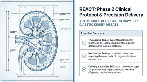
For decades, the standard of care for Type 2 Diabetic Kidney Disease (DKD) has followed a predictable and disheartening trajectory. As the largest patient demographic facing renal failure globally, these individuals are often relegated to a managed decline. Clinicians monitor the slow erosion of function until the system reaches a breaking point, at which stage dialysis—a life-altering and exhausting intervention—becomes the only path to survival.However, a new paradigm is emerging: Renal Autologous Cell Therapy (REACT). Rather than merely managing the symptoms of a failing system, REACT represents a fundamental shift toward active architectural repair. By executing localized, cellular-level interventions, this technology moves beyond the traditional model of systemic failure. It is no longer about slowing the slide; it is about recalibrating the kidney’s own infrastructure to preserve its life-sustaining work.
2. The “eGFR 15” Trap: Why Preservation is the New Cure
In the traditional clinical pathway, once a patient’s estimated Glomerular Filtration Rate (eGFR) drops below 15, they are funneled toward dialysis initiation. This transition often correlates with a sharp decrease in life expectancy. The REACT pathway proposes a different strategy: the preservation of “Residual Kidney Function” (RKF).The REACT strategy targets patients in a specific “sweet spot”—Chronic Kidney Disease (CKD) Stages 3a–4, where eGFR remains between 20 and 50. By intervening before the architecture has completely collapsed, the therapy aims to maintain the kidney’s biological baseline.”If we maintain even a fractional eGFR, the patient’s predicted life expectancy improves dramatically over total renal failure.”The most profound insight from recent clinical dossiers is that maintaining an eGFR even in the 10–15 range results in a prolonged prognosis regardless of whether the patient eventually requires dialysis. In this new model, preservation is not a delay of the inevitable; it is a clinical victory that fundamentally alters the patient’s survival map.
3. The Autologous Loop: Turning Your Own Cells into Your Best Medicine
REACT utilizes a 100% Autologous Protocol, meaning the therapeutic agent is derived entirely from the patient’s own native tissue. This eliminates the immunological risks of donor-based therapies and the “one-size-fits-all” limitations of standardized drugs. The process follows a rigorous three-step cycle:
- Precision Tissue Extraction (Biopsy): Using real-time CT guidance, clinicians perform a percutaneous harvest of native renal tissue. This requires a precise depth penetration of 1.5cm to 1.8cm to secure viable functional tissue from the renal cortex.
- Cellular Processing & Selection: The extracted core is immediately stabilized and sent to a GMP (Good Manufacturing Practice) expansion facility. The tissue undergoes enzymatic digestion and a strict 6-week cultivation cycle. During this phase, density-gradient centrifugation is employed to select specific, viable cellular subpopulations critical for regeneration.
- Targeted Reinjection: Once the cells are formulated into a protective gelatin-based hydrogel matrix, they are delivered back into the patient’s renal cortex. This intervention must be administered within a strict 72-hour clinical window post-formulation to ensure maximum cell viability.
4. Beyond the Human Touch: The Mandate for Sub-Centimeter Precision
The primary technical bottleneck in renal therapy is the unforgiving anatomy of the organ. The target zone for repair—the renal cortex—is strictly less than 5mm deep. The total thickness of the outer layers (the renal capsule and cortex combined) is a mere 7mm to 10mm.Traditional free-hand surgery suffers from a “Surgical Blind Spot.” The human hand cannot physically feel a 0.5cm anatomical variance deep inside the body, leading to inconsistent deposition. REACT replaces “approximated surgery” with “precision engineering.”The protocol utilizes a sophisticated needle-in-needle system: a 20G outer co-axial needle acts as a stable sheath, while a 25G inner delivery needle precisely deposits the cells. Guided by computational mathematical modeling and real-time CT monitoring, clinicians calculate exact coordinates to distribute the cells across multiple sites. By employing a “pull-and-inject” method—where the inner needle is retracted incrementally—the cells are deposited safely into the fragile interstitial space without causing a “pressure blowout,” ensuring they reach the diverse microenvironments necessary to trigger regenerative signaling.
5. Personalized Math: Why Your Dose is Calculated via MRI
Unlike standardized pharmaceuticals, REACT is a personalized biological intervention. There is no “standard dose”; instead, every patient receives a unique cellular volume derived through individualized mathematical modeling.Before the reinjection protocol begins, an MRI volumetric image analysis is performed to determine the exact mass of the patient’s kidney. This is essential because organ mass varies significantly; the baseline for males typically ranges from 120g to 170g, while the female average ranges from 115g to 155g.
Individualized Dosing Formula Volume/Mass of Kidney * Targeted Cells per gram (3 * 10^6) = Individualized Syringe Dose
6. The Bio-Logic: Repairing the Filtration Barrier
At the heart of kidney function is the Glomerular Basement Membrane (GBM). This is a highly specialized porous barrier composed of a matrix of Type 4 Collagen and Laminins , supported by podocytes that create ultra-fine filtration slits of 80-100nm. When this architectural matrix is damaged, the kidney loses its ability to filter plasma, and eGFR plummets.The biological mechanism of REACT is modeled after neural progenitor cells. In the nervous system, progenitor cells differentiate into Oligodendrocytes for structural myelin repair or Astrocytes for nutrition and support. Similarly, the density-gradient selected cells injected into the kidney respond to local chemical markers. Once deposited, they differentiate to repair the specific damaged filtration architecture of the GBM. Restoring this structural architectural integrity is the “Holy Grail” of preserving renal function.
7. Conclusion: A Shift in the Medical Compass
The transition from managing decline to executing localized repair rests on three pillars:
- Precise Identification: Utilizing a strict diagnostic matrix to identify patients in the 20-50 eGFR “sweet spot” (Ages 30-80, HbA1c < 10, BMI < 45).
- Autologous Regeneration: Leveraging the patient’s own native tissue through GMP-grade enzymatic digestion and subpopulation selection.
- Spatial Accuracy: Eliminating human variance through CT-guided, multi-site deposition and sub-centimeter coordinate calculation.As we master the ability to engineer the body to repair its own infrastructure with sub-centimeter precision, the nature of our medical mission changes. When we can target the renal cortex with the accuracy of a guided missile, does “chronic disease” start to look like an engineering problem we’ve finally learned to solve?
Summary Video
Summary File